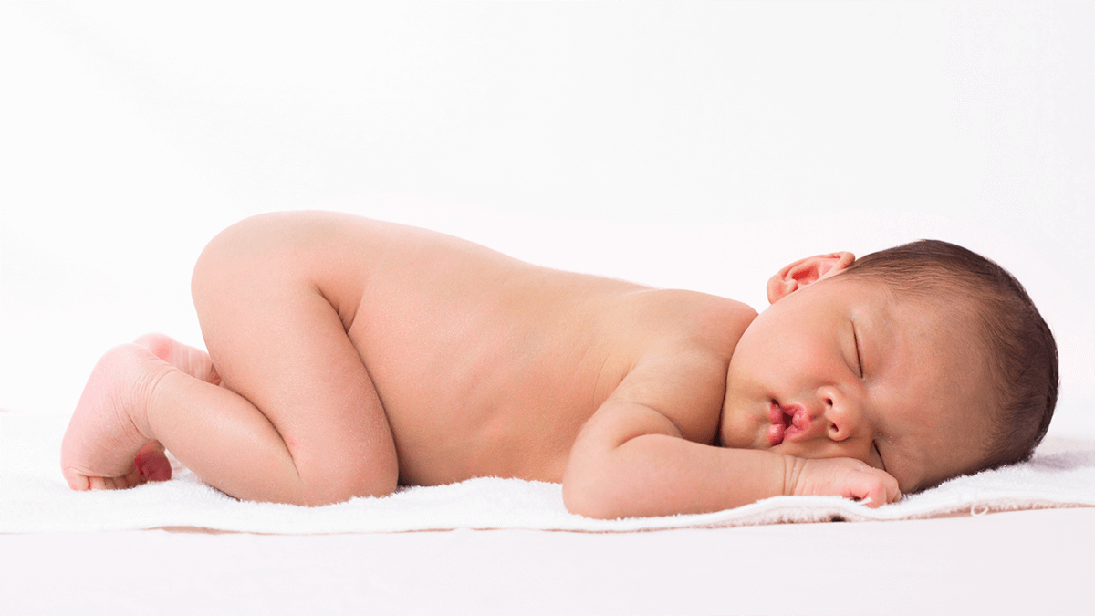
Can Sonograms Detect Cancer?

Can Sonograms Detect Cancer?

A cancer diagnosis can be daunting, and understanding the available tools is crucial. Ultrasounds, often referred to as sonograms, are a common imaging technique used for various medical purposes. But can they actually detect cancer? This guide is here to explain every detail related to cancer detection and sonograms. Let’s look into it together!

What is Ultrasound Technology?

Mammography employs ultrasonic waves to produce images of internal organs and most body tissues. These sound waves are emitted from a portable transducer. When waves pass through your body, they reflect in various tissues when they meet different ones. These returning echoes are then picked up by the transducer, which gets rid of its surrounding tissue and converts them into electrical signals. A computer will analytically study these signals and translate them into the visual form you see in the sonograms.

Sonograms and Cancer Detection

While sonograms can’t definitively diagnose cancer on their own, they play a vital role in the detection process. Here’s why:

  • Visualizing Abnormalities: It can also demonstrate the presence or absence of lumps, masses, nodularity, or an increase in size, hardness, or softness of the breast tissue, which may be an early sign of cancer.
  • Guiding Biopsies: Doctors can rely on them to take tissue samples – biopsies – from some parts of the body that look cancerous with the help of a microscope to determine cancer.
  • Monitoring Treatment: Another application of ultrasound imaging is evaluating cancer treatment by checking the size and response of tumors.

Limitations of Sonograms in Cancer Detection

Despite their benefits, sonograms have limitations in cancer detection:

  • Resolution: Sonograms are not as detailed as some of the other imaging techniques, such as CT scans or MRI. They might fail to detect a tumor if it is too small or situated in areas that cannot effectively respond to sound waves, such as the lungs.
  • Distinguishing Benign from Malignant: Due to the limitations of sonograms’ distinctions, radiologists can’t be certain whether the growth is benign (non-cancerous) or malignant (cancerous). On most occasions, tissue biopsies are needed to establish a diagnosis.

When are Sonograms Used in Cancer Detection?

Ultrasound is often used to examine organs readily accessible with sound waves, such as:

  • Breast: This may be useful in determining areas in the breast that may have formed lumps, cysts, or any other irregularities that need a biopsy.
  • Thyroid: Enables to see nodules themselves or changes in the size of the thyroid gland.
  • Liver: This one is able to identify conditions such as enlarged livers, tumors or any other irregularities present in one’s liver.
  • Pelvis: This is used to study the shape and size of the uterus, ovaries, and other pelvic organs for signs of lesions.

Peace of Mind During Pregnancy: Ultrasound for You!

While sonograms can’t detect cancer, 4Baby Ultrasound provides a safe and comfortable environment for your prenatal ultrasounds. See your baby in stunning 2D, 3D, 4D, or even 5D detail and bond with your little one early on. Schedule your worry-free pregnancy ultrasound at 4Baby Ultrasound today!

Share This Post

Logo

Our 4 Baby ultrasound and sonogram sessions allow you to see your unborn baby in 2d, 3d, and 4d live motion with a 3D/4D/HD ultrasound at our locations in Texas, serving Dallas, Fort Worth, Waxahachie, Ennis, Grapevine, Frisco, Flower Mound and Tyler Texas as well. 

More To Explore

Baby Walkers Good for Babies

Most parents may opt to purchase a baby walker to assist their children in walking. But are baby walkers really beneficial to babies in any

How Long Can You Leave a Newborn Alone?

In the early days, newborns are completely dependent and require constant care. Many parents wonder when it’s safe to leave their baby alone and for

Request A

Consultation