Why Every Parent Should Get 3D/4D Ultrasound Baby Pictures
Pregnancy is an exciting journey that is filled with joy and a little bit of nervousness. Every parent wonders what their baby will look like,

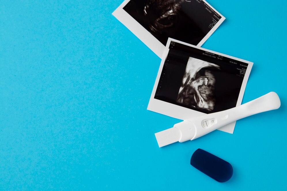
3d Sonogram: A 3D (three-dimensional) ultrasound scan shows detailed pictures of your baby. Doctors recognize these pictures visually by the orange or golden color and details of the baby’s face; nose, eyes, mouth, etc. Therefore, a 3d sonogram is helpful to look at the heart and other internal organs. As an outcome, some clinics use 3D ultrasound scans, but only when medically necessary.
With multi-dimensional ultrasound imaging, doctors can fully understand the spatial relationships of the anatomical parts they are looking at and better assess a particular condition, like:
A cleft lip is one example. In this condition, the tissue from each head side does not join entirely at the lip, leaving a gap. Although a 2D scan may indicate the issue, a 3d sonogram Dallas offers a better view of the range of the split.
3D ultrasounds provide an image of the placenta, but they can also help assess its vascularization, volume, and blood flow depending on the technology used. A 3D sonogram can also look at the placenta’s placement in the womb, particularly in placenta accreta, where the placenta grows deeply into the uterine wall.
A 3d sonogram Dallas makes it easier for doctors to interpret scans of the fetal heart anatomy. But it depends on technology usage; a 3D/4D ultrasound baby ultrasound can even explore how the heart correlates with the pots and structures.

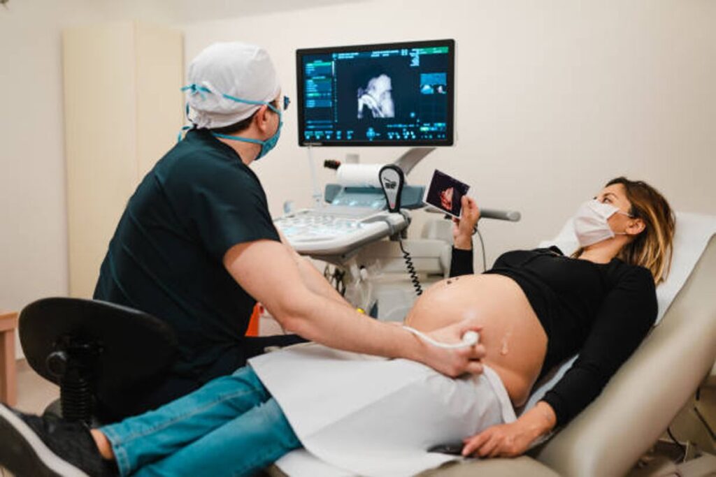
Our 4 Baby ultrasound and sonogram sessions allow you to see your unborn baby in 2d, 3d, and 4d live motion with a 3D/4D/HD ultrasound at our locations in Texas, serving Dallas, Fort Worth, Waxahachie, Ennis, Grapevine, Frisco, Flower Mound and Tyler Texas as well.

Pregnancy is an exciting journey that is filled with joy and a little bit of nervousness. Every parent wonders what their baby will look like,

Shopping for a baby shower gift can be tricky. With so many options, how do you know what the parents really need? A gift card
2025 © 4babyultrasound All rights reserved | Powered by 4Baby Ultrasound & Photography Boutique